Unlock the Secrets: What Inner Cell Mass Becomes After?

Understanding the developmental biology of early embryos requires grasping a fundamental question: what does the inner cell mass become? The inner cell mass, a critical cluster of cells, gives rise to the epiblast and the primitive endoderm; these early structures will eventually form the entire body of the organism and its supporting tissues. Researchers at institutions like the Carnegie Institution for Science are using advanced microscopy techniques and cellular assays to study these developmental processes. This exploration seeks to unravel the intricate mechanisms determining cellular fates and provides insights into how the inner cell mass becomes the complex organism.
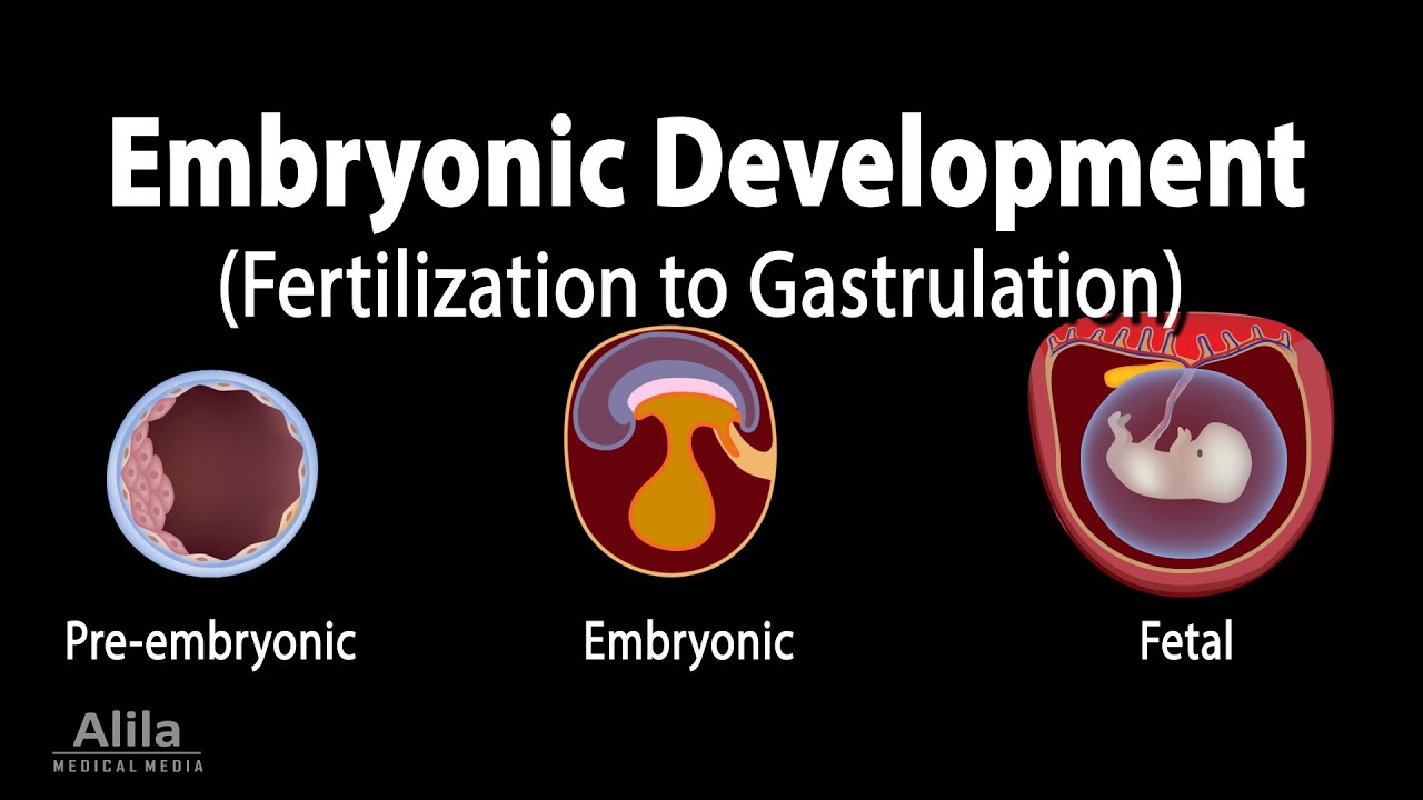
Image taken from the YouTube channel Alila Medical Media , from the video titled Embryology: from Fertilization to Gastrulation, Animation .
The journey from a single fertilized egg to a fully formed organism is one of the most remarkable processes in biology. Central to this transformation is a small group of cells nestled within the early embryo, known as the Inner Cell Mass (ICM). Understanding the ICM, its origins, and its developmental potential, is paramount to unraveling the mysteries of life itself.
Defining the Inner Cell Mass: The Seed of Development
The ICM, also referred to as the embryoblast, is a cluster of cells located inside the blastocyst, an early stage of the developing embryo. The blastocyst, a hollow sphere, consists of two primary cell types: the trophectoderm, which will eventually form the placenta, and the ICM.
The ICM's strategic position and unique cellular properties mark it as the origin of the entire organism. It is from these few cells that all the tissues and organs of the developing body will arise.
Why the ICM Matters: Implications for Development and Medicine
The significance of the ICM extends far beyond basic developmental biology. By understanding the intricate mechanisms that govern its fate, we can gain valuable insights into a range of biological processes. These mechanisms have vast implications for regenerative medicine and disease modeling.
Understanding the ICM holds keys to:
- Developmental biology: Illuminating the fundamental processes that shape the body plan.
- Regenerative medicine: Harnessing the ICM's potential to repair damaged tissues and organs.
- Disease modeling: Creating cellular models of diseases to study their causes and develop new treatments.
- Treating infertility: Improving assisted reproductive technologies.
The knowledge gained from studying the ICM has the potential to revolutionize how we approach human health and disease.
Roadmap: Charting the ICM's Developmental Course
To fully appreciate the ICM's role, we must trace its developmental journey. The sections that follow will explore the key stages of this process:
We will begin by examining the ICM within its blastocyst home, distinguishing it from the trophectoderm. Next, we will delve into cellular differentiation, exploring how the ICM cells transition from a pluripotent state to specialized cell types. We will then examine gastrulation and the formation of the three germ layers – ectoderm, mesoderm, and endoderm. We will continue to see how they lay the foundation for all tissues and organs. From there, we will explore organogenesis, the intricate process by which organs develop from these germ layers. Finally, we will discuss the close relationship between the ICM and Embryonic Stem Cells (ESCs), and their potential in regenerative medicine, while addressing the ethical considerations surrounding this research.
By carefully examining each of these stages, we will gain a comprehensive understanding of the Inner Cell Mass and its enduring legacy in shaping the organism.
The journey from a single fertilized egg to a fully formed organism is one of the most remarkable processes in biology. Central to this transformation is a small group of cells nestled within the early embryo, known as the Inner Cell Mass (ICM). Understanding the ICM, its origins, and its developmental potential, is paramount to unraveling the mysteries of life itself.
As we consider the ICM's pivotal role, it's crucial to understand the context in which it exists. The ICM isn't simply floating in space; it resides within a structure called the blastocyst, a critical stage in early embryonic development.
The ICM's Residence: The Blastocyst Stage
The blastocyst is an early-stage embryo characterized by its distinctive structure: a fluid-filled cavity surrounded by an outer layer of cells, with a cluster of cells tucked inside.
Think of it like a miniature cellular globe, with two distinct populations destined for vastly different fates.
Anatomy of the Blastocyst
The blastocyst is comprised of two primary cell types:
-
Trophectoderm: The outer layer of cells forms the wall of the blastocyst. It is responsible for implanting the embryo into the uterine wall and eventually forming the placenta.
-
Inner Cell Mass (ICM): This compact cluster of cells resides inside the blastocyst cavity. As mentioned earlier, it is the source of all the tissues and organs of the developing embryo.
Imagine a hollow ball (the blastocyst) with cells lining the outside (trophectoderm) and a small clump of cells (the ICM) attached to the inside wall.
Visualizing this structure is essential for grasping the ICM's strategic position and its subsequent role in development.
Trophectoderm vs. ICM: A Tale of Two Fates
While both the trophectoderm and the ICM are essential for successful development, their ultimate destinies differ dramatically.
The trophectoderm, destined to form the placenta, is critical for nourishing and supporting the developing embryo.
However, it is the ICM, with its remarkable capacity for self-renewal and differentiation, that truly holds the keys to creating a new organism.
The trophectoderm is a supporting player, paving the way for the ICM's grand performance.
This fundamental distinction – one destined for support, the other for building – highlights the exquisite orchestration of early embryonic development.
Pluripotency: The Hallmark of ICM Cells
Perhaps the most remarkable characteristic of ICM cells is their pluripotency.
This term signifies their ability to differentiate into any cell type in the body, whether it be a neuron, a muscle cell, or a skin cell.
It is this "blank slate" nature that makes the ICM so powerful, so full of potential.
Think of them as master builders, capable of constructing any structure from a single blueprint.
This unique property allows the ICM to give rise to all three germ layers (ectoderm, mesoderm, and endoderm) during gastrulation, which will later form the foundation for all tissues and organs in the body.
Pluripotency is the defining feature that sets the ICM apart and makes it such a focus of scientific inquiry.
The pluripotent nature of the ICM has led to another groundbreaking discovery: Embryonic Stem Cells (ESCs).
ESCs are derived directly from the ICM of early embryos.
Crucially, they retain the ICM's ability to differentiate into any cell type, making them invaluable tools for research and potential therapies.
Scientists can grow ESCs in the lab and, under the right conditions, coax them to become specific cell types.
This opens up exciting possibilities for regenerative medicine, where ESCs could be used to repair damaged tissues or replace organs.
ESCs represent the ICM's legacy extended – a powerful tool derived from the early embryo with the potential to revolutionize medicine.
As we consider the remarkable capabilities of the ICM, we must also investigate how these initially identical cells embark on divergent paths to create the myriad tissues and organs of the body. This transformative process, known as cellular differentiation, is the linchpin of embryonic development.
Differentiation Unveiled: The ICM's Journey to Specialization
Cellular differentiation is the cornerstone of embryonic development, driving the transition from a cluster of identical, pluripotent cells to the diverse array of specialized cells that comprise a complete organism. Understanding this process is essential to grasping how the ICM, with its seemingly unlimited potential, ultimately gives rise to the complex architecture of the body.
Defining Differentiation in Embryonic Development
At its core, differentiation is the process by which a less specialized cell becomes a more specialized cell type.
In the context of the developing embryo, this means that the ICM cells, initially capable of becoming any cell in the body, gradually restrict their developmental potential and adopt specific identities.
This transition involves a cascade of molecular events, ultimately leading to distinct changes in cell structure, function, and gene expression.
The Symphony of Influence: Factors Governing Differentiation
The differentiation of ICM cells isn't a spontaneous or random event. It's a carefully orchestrated process, driven by a complex interplay of internal and external factors. These factors act as cues, guiding cells down specific developmental pathways.
Signaling Molecules: The Language of Cellular Communication
Signaling molecules are key players in differentiation.
These molecules, often proteins or peptides, are secreted by cells within the embryo and act as messengers.
They bind to receptors on the surface of other cells, triggering intracellular signaling cascades that ultimately alter gene expression.
For example, growth factors, morphogens, and cytokines can all influence the fate of ICM cells by activating or inhibiting specific signaling pathways.
The Epigenetic Landscape: Gene Expression Control
Epigenetics also plays a key role in differentiation. While all cells in the body contain the same genetic information, the specific genes that are turned on or off vary dramatically between different cell types. Epigenetic modifications, such as DNA methylation and histone modification, control which genes are accessible for transcription. These modifications can be influenced by signaling molecules and other environmental factors, further fine-tuning the differentiation process.
Internal Genetic Programs
Cells also have intrinsic genetic programs that guide their differentiation.
These programs are encoded in the cell's DNA and are activated in response to specific signals.
Transcription factors, proteins that bind to DNA and regulate gene expression, are crucial components of these programs.
Commitment to Lineage: From Potential to Reality
The journey of an ICM cell to becoming a specialized cell type is often described as a process of progressive restriction.
Initially, ICM cells are pluripotent, meaning they can become any cell in the body. As development proceeds, these cells gradually lose their ability to differentiate into certain cell types, becoming more and more committed to a particular lineage.
Think of it like a branching path, where cells make decisions at each fork, narrowing down their options until they reach their final destination.
This commitment is often irreversible, meaning that once a cell has committed to a particular lineage, it cannot easily revert to its pluripotent state.
This intricate process ensures that the right types of cells are created in the right places at the right times, ultimately giving rise to a functional and well-organized organism.
As cells differentiate, they begin to express specific genes and take on distinct characteristics. However, the organization of these differentiating cells is just as crucial as their individual identities. This coordinated choreography culminates in the formation of the three primary germ layers, which act as the foundational blueprint for the entire body.
The Three Germ Layers: The Foundation of Body Plan Development
Gastrulation marks a pivotal moment in embryonic development: the formation of the three primary germ layers: ectoderm, mesoderm, and endoderm. These layers are not merely collections of cells; they are the foundational tissues from which all the organs and structures of the fully formed organism will arise. Understanding their formation and fate is crucial to understanding the entire body plan.
Gastrulation: Orchestrating the Formation of the Germ Layers
Gastrulation is the dynamic process by which the relatively simple, hollow blastocyst undergoes a dramatic reorganization. It is a carefully orchestrated cellular dance that establishes the three germ layers.
Think of it as the embryo's initial attempt at architectural design, laying the groundwork for future construction.
The cells of the ICM migrate and rearrange themselves, creating distinct layers that will give rise to specific tissues and organs. This process is driven by a complex interplay of signaling molecules and cell-cell interactions.
The Ectoderm: The Outer Layer of Protection and Connection
The ectoderm is the outermost germ layer. It is the precursor to the structures that interface with the external world: the skin and the nervous system.
The Skin: Our Protective Barrier
The ectoderm gives rise to the epidermis, the outer layer of our skin, providing a protective barrier against the environment.
It also forms structures like hair, nails, and sweat glands, all essential for protection and regulation.
The Nervous System: The Body's Control Center
Perhaps most remarkably, the ectoderm is the source of the entire nervous system, including the brain, spinal cord, and peripheral nerves.
This complex network allows us to perceive, process, and respond to the world around us.
The Mesoderm: The Foundation of Structure and Movement
The mesoderm is the middle germ layer, responsible for the body's structural components, movement systems, and circulatory system.
Muscles and Bones: Enabling Movement and Support
The mesoderm gives rise to muscles, enabling movement, and the skeletal system providing support and structure.
The Circulatory System: Delivering Life
Crucially, the mesoderm also forms the heart and blood vessels. This complex network ensures that every cell in the body receives the oxygen and nutrients it needs to survive and function.
The Endoderm: Lining the Internal World
The endoderm is the innermost germ layer.
It forms the lining of the digestive and respiratory tracts, as well as key organs like the liver and pancreas.
The Digestive and Respiratory Systems: Essential for Survival
The endoderm forms the epithelial lining of the digestive tract and respiratory tract, which are responsible for nutrient absorption and gas exchange, respectively.
The Liver and Pancreas: Vital Metabolic Organs
The liver and pancreas, essential for metabolism and digestion, also originate from the endoderm.
The Germ Layers as the Blueprint for the Body
The three germ layers are not merely transient structures; they are the foundational blueprint upon which the entire body is built. Each layer is destined to give rise to a specific set of tissues and organs.
This precise allocation of developmental potential ensures that the body develops in a coordinated and organized fashion.
Without the proper formation and differentiation of the germ layers, the development of a functional organism would be impossible.
Gastrulation lays the groundwork, but the true architectural marvel of development unfolds during organogenesis. The simple, elegant arrangement of the three germ layers becomes the foundation upon which intricate and specialized organs are constructed. This is where the blueprint of life translates into tangible form, transforming sheets of cells into the functional components of a complete organism.
Organogenesis: Sculpting Organs from Germ Layer Foundations
Organogenesis is the remarkably complex developmental process where the three primary germ layers – ectoderm, mesoderm, and endoderm – differentiate and interact to form the various organs of the body. This orchestrated sequence of events involves cell proliferation, migration, differentiation, and programmed cell death (apoptosis), all working in concert to shape functional organs. It's not merely about assembling cells; it's about sculpting them into intricate structures with highly specific functions.
The Germ Layer Lineage: A Blueprint for Organ Development
Each germ layer possesses a distinct developmental fate, giving rise to a specific set of organs and tissues:
-
Ectoderm: This outer layer is the precursor to structures that interface with the outside world. The neural tube, the embryonic precursor to the central nervous system (brain and spinal cord), arises from the ectoderm. Additionally, the ectoderm gives rise to the epidermis, including skin, hair, and nails.
-
Mesoderm: Occupying the middle ground, the mesoderm is responsible for the body's structural and supportive elements. The heart, the first functional organ to develop, originates from the mesoderm. In addition, the mesoderm gives rise to muscle tissue, bone, blood cells, kidneys, and the reproductive system.
-
Endoderm: As the innermost layer, the endoderm forms the linings of the digestive and respiratory tracts, as well as associated organs. The liver and pancreas, vital for metabolic function, develop from the endoderm. The endoderm also contributes to the formation of the thyroid and thymus glands.
Orchestrating the Symphony: Cellular Signaling and Genetic Regulation
Organogenesis isn't a haphazard process; it's a meticulously controlled symphony of cellular signaling and genetic regulation. Cells communicate with each other through a variety of signaling pathways, including:
- Growth Factors: These proteins stimulate cell proliferation and differentiation.
- Morphogens: These signaling molecules act as positional cues, instructing cells about their location and fate within the developing organ.
- Cell Adhesion Molecules: These proteins mediate cell-cell interactions, ensuring that cells adhere to the correct neighbors and form organized tissues.
These signaling pathways activate specific genes within cells, leading to changes in cell shape, behavior, and ultimately, the formation of functional tissues and organs.
Tissues as Building Blocks: Form and Function in Organ Development
Organs are rarely composed of a single cell type. Instead, they are complex assemblies of different tissues, each contributing to the organ's overall structure and function. For example, the heart contains:
- Muscle tissue (myocardium): Generates the force for pumping blood.
- Connective tissue: Provides structural support and elasticity.
- Epithelial tissue (endocardium and pericardium): Lines the inner and outer surfaces of the heart.
- Nervous tissue: Regulates heart rate and contraction strength.
The precise arrangement and interaction of these tissues are essential for the heart to function properly. Similarly, other organs rely on the coordinated action of multiple tissue types to perform their specialized roles.
The ICM and Embryonic Stem Cell Research: A Bridge to Regenerative Medicine
The inner cell mass (ICM) is not just a fleeting stage in embryonic development; it's a foundational source of embryonic stem cells (ESCs), holding immense promise for regenerative medicine and disease modeling. These cells, with their unique ability to differentiate into virtually any cell type in the body, represent a potential revolution in how we treat injuries and illnesses.
The Direct Lineage: From ICM to ESCs
ESCs are, in essence, the cultured descendants of the ICM. When isolated from the blastocyst, ICM cells can be grown in vitro under specific conditions that maintain their pluripotency. These self-renewing cells provide a continuous source of undifferentiated material.
This direct lineage is what makes ESCs so valuable. They retain the ICM’s inherent developmental potential. It offers researchers an unprecedented opportunity to study early human development and differentiation processes in a controlled laboratory setting.
ESCs: A Powerful Tool for Regenerative Medicine
The true potential of ESCs lies in their ability to generate functional cells, tissues, and even potentially whole organs, to replace those damaged by disease or injury.
Imagine a future where damaged heart muscle is replaced with new, healthy cells derived from ESCs, effectively reversing the effects of a heart attack. Or where patients with spinal cord injuries can regain lost function through the transplantation of ESC-derived neural cells. This is the promise of regenerative medicine.
Here are some of the specific areas where ESCs hold tremendous promise:
- Treating Degenerative Diseases: ESCs could be used to generate dopamine-producing neurons to treat Parkinson's disease or insulin-producing beta cells to treat type 1 diabetes.
- Repairing Damaged Tissues: ESCs could be differentiated into cardiomyocytes to repair damaged heart tissue after a heart attack or into skin cells to heal severe burns.
- Drug Discovery and Toxicity Testing: ESC-derived cells can be used to create more accurate and relevant models for testing the efficacy and safety of new drugs.
- Disease Modeling: ESCs can be used to create disease-specific cell lines, allowing researchers to study the mechanisms of disease and develop new therapies.
The Ethical Landscape of ESC Research
The promise of ESC research is undeniable, but it is intertwined with significant ethical considerations. The primary concern revolves around the source of ESCs, which, in most cases, involves the destruction of human embryos.
This raises fundamental questions about the moral status of the embryo and the permissibility of using embryos for research purposes, even if it could lead to life-saving treatments.
Navigating the Ethical Dilemmas
The ethical debate surrounding ESC research is complex and multifaceted, with diverse perspectives rooted in different moral, religious, and philosophical beliefs.
Some argue that the potential benefits of ESC research outweigh the moral concerns, emphasizing the potential to alleviate suffering and save lives. Others maintain that the destruction of human embryos is inherently wrong, regardless of the potential benefits.
Finding a balance between these competing values is crucial. Many researchers and policymakers advocate for strict ethical guidelines and oversight to ensure that ESC research is conducted responsibly and ethically.
Alternative Approaches: Induced Pluripotent Stem Cells (iPSCs)
It's important to note that advancements in stem cell technology have provided alternative approaches that circumvent the need to use embryos.
Induced pluripotent stem cells (iPSCs), for example, are generated by reprogramming adult cells back to a pluripotent state, similar to ESCs. This breakthrough has opened up new avenues for regenerative medicine while also addressing some of the ethical concerns associated with ESC research.
While iPSCs offer a promising alternative, ESC research remains valuable for understanding the fundamental biology of pluripotency and differentiation, and for developing new stem cell-based therapies. The journey from the ICM to clinical applications is ongoing, requiring continuous scientific innovation and thoughtful ethical reflection.
Video: Unlock the Secrets: What Inner Cell Mass Becomes After?

FAQs: Inner Cell Mass Fate
These frequently asked questions clarify what happens to the inner cell mass (ICM) after its initial formation during embryonic development.
What is the inner cell mass's main role?
The inner cell mass (ICM) is crucial for forming the entire body of the developing organism. It's a cluster of cells within the blastocyst that ultimately gives rise to all the tissues and organs. Without a properly functioning ICM, normal development cannot proceed.
Specifically, what does the inner cell mass become?
The inner cell mass differentiates into two main layers: the epiblast and the hypoblast. The epiblast is responsible for forming the three primary germ layers (ectoderm, mesoderm, and endoderm), which give rise to all the structures of the embryo. So, the inner cell mass becomes the basis for the entire fetus.
How does the hypoblast relate to what the inner cell mass becomes?
While the epiblast becomes the embryo, the hypoblast forms extraembryonic tissues, specifically the yolk sac. The yolk sac provides nourishment to the early embryo. Although not directly becoming part of the fetus, the hypoblast plays a vital supporting role.
Is the inner cell mass the same as a stem cell?
The inner cell mass contains pluripotent stem cells. This means they have the potential to differentiate into any cell type in the body. Researchers study what the inner cell mass becomes to understand and harness the power of these cells for regenerative medicine.